

Breast Cancer Basics: Third In A Four-Part Series
When You Need A Breast Biopsy; Dr. Barry Roseman Offers Health Advice

In this article, the third in a series about breast cancer, we focus on breast biopsies.
To begin with, let me define the word biopsy. A breast biopsy is the removal of tissue from the breast of a woman (or man) to be examined for the presence of a disease – most commonly to determine if breast cancer is present.
What would lead a physician to recommend a biopsy? Typically, a biopsy is done because of new or “suspicious” findings on breast imaging studies such as a mammogram, breast ultrasound or breast MRI (magnetic resonance imaging) scan. Occasionally new breast lumps are found on a breast exam, and while the next step to determine the nature of such lumps is to perform breast imaging, this may also lead to recommendations for a biopsy.
Biopsies are “image guided,” that is, performed with diagnostic radiology imaging to ensure that the tissue being removed corresponds to the radiologic abnormality. We want to see the abnormal finding while doing the biopsy, to be sure that we are sampling the precise area in question.
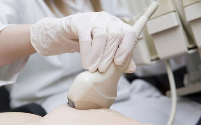
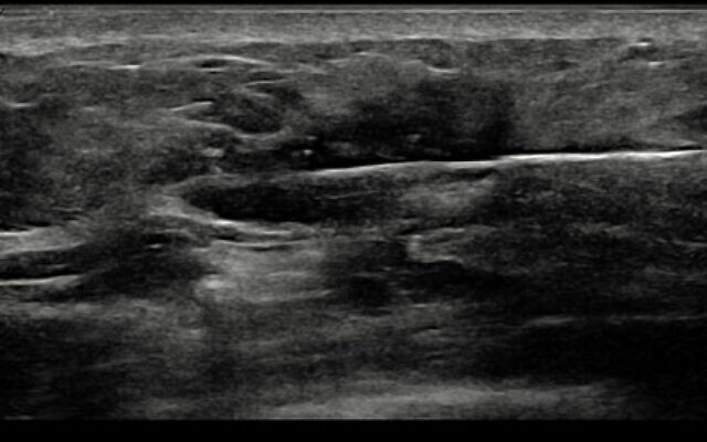
If there is a breast lump or mass present, it is commonly visible with ultrasound, and in this case, ultrasound can be used to guide the biopsy. If the finding is only seen on a mammogram or MRI scan, then that same imaging device is used to “guide” the exact biopsy location.
Ultrasound biopsies can be performed in the office. After using a small amount of local anesthetic in the skin, a small needle is inserted through the skin that removes fragments of breast tissue. Lymph nodes can be sampled under ultrasound guidance in a similar fashion.
Mammogram-guided biopsies are typically done in a breast center, and MRI guided biopsies are even more elaborate, as they need to be done by a team while you are in the MRI scanner.
For several reasons, a very small titanium biopsy “clip” or “marker” is typically left at the site of the biopsy. This identifies the site on future imaging so that radiologists will know that a biopsy was done, and to ensure that the correct area was sampled. The clips are also useful if there is additional surgery, to precisely locate the site that was biopsied. Think of it as similar to leaving cookie crumbs, as a trail back to the original location.

The tissue samples removed are immediately sent to a pathology lab where they are studied, and a report is created to describe the appearance of the tissue under the microscope and establish the final diagnosis. When breast cancer is present, additional stains are used to determine details of the tumor biology, that is, what factors are contributing to tumor growth and what treatments will be appropriate and effective.
Typically, the biopsy results will be conclusive. The pathologists are experienced enough that they will not miss cancer, nor call something cancer by mistake. The only thing to keep in mind is that a negative biopsy does not completely rule out cancer. Think of it this way: you reach in your purse and cannot find your credit card. Does that mean it is not there? Probably, but if you empty the purse out completely and look in every nook and cranny, and still don’t find it – now you’re sure.
The same goes with sampling breast tissue. The larger the sample without cancer, the surer we can be, but without removing the entire abnormality surgically we can never be 100 percent sure.
Which leads to the subject of a surgical biopsy. In the past, before the development of sophisticated mammogram and ultrasound imaging, this was the only way to do a biopsy. Open surgical biopsies are rarely done now for breast disease, as we prefer to avoid doing an operation just to diagnose a problem.
However, sometimes surgery is required to obtain enough tissue to make a firm pathology diagnosis. In this case, a surgeon takes you to a sterile operating room, administers local or general anesthesia, makes an incision in the skin, then completely removes the abnormal mass or radiologic abnormality. When the results are benign, you can be safely monitored or observed. When breast cancer is found, additional surgery is often necessary to definitively remove the tumor, and sometimes sample lymph nodes.
If a Breast Biopsy is recommended for you, do not worry, just have it done. Biopsies today can be simple procedures with mild to little pain, and you always need to know what is going on in your breasts (and your body). Remember that the earlier Breast Cancer is diagnosed, the less treatment will be necessary, and the better the outcome will be!
My next and final article will deal with breast cancer treatment.
If you have questions or wish to contact me, please call or email me and I will be happy to respond.
Dr. Barry Roseman
Advanced Breast Care / Surgical
Oncology of North Georgia, Inc
www.advanced-breastcare.com
404-841-6262
barry.roseman@gmail.com
Paid Content by Advanced Breast Care
comments